Diagram Cross Section Of A Bone / Bone Cross Section High Res Stock Images Shutterstock - Explaned distal and proximal epiphysis.. The cross section of a rectangular pyramid is a rectangle. The diagram of a long bone could become your choice when making about bone. Each system contains for a bone tissue engineering scaffold to be successful, it must be highly porous, osteoconductive, biodegradable, biocompatible, mechanically. For example, to read this diagram literally, since the cartilage can be seen inside the cutaway section of bone, it incorrectly indicates that the cartilage in fact goes through the bone structure, rather than just being found around the bone end. Select from premium cross section of bone images of the highest quality.
From wikimedia commons, the free media repository. Metaphseal region on the left, diaphyseal region on the right. There are trabeculae in spongy bone which gives its sponge like appearance. Jump to navigation jump to search. Diagram with articular cartilage, marrow, spongy bone, medullary cavity, endosteum, diaphysis, and periosteum.
Cross Section Bone Human High Resolution Stock Photography And Images Alamy from c8.alamy.com Bone is found in the shafts of long bone and consists of various cylindrical units named as haversian system 47. Human respiratory system anatomical line style artistic vector illustration, medical education cross section diagram. There are trabeculae in spongy bone which gives its sponge like appearance. How to draw the diagram of cross section of a leaf class x. Related posts of cross section of human bone diagram. Cross section of a bone. Bone marrow is the soft, highly vascular and flexible connective tissue within bone cavities which serve as the primary site of new blood cell production or bone marrow is the primary source of pluripotent stem cells that give rise to all hemopoietic cells (blood cells) including lymphocytes. (b) in this micrograph of the osteon, you can clearly see the concentric lamellae and central canals.
The cross section of this circular cylinder is a circle.
There are trabeculae in spongy bone which gives its sponge like appearance. There are trabeculae in spongy bone which gives its sponge like appearance. Some descriptions for confusing partsomit number 13 in the picture. As a part of the. Select from premium cross section of bone images of the highest quality. Cross sections are usually parallel to the base like above, but can be in any direction. Diagram of a cross section of the coiled cochlea. □ the osteon consists of a system of bony lamellae arranged concentrically around a canal, which is called haversian canal. As shown in figure 2. Explaned distal and proximal epiphysis. Metaphseal region on the left, diaphyseal region on the right. From wikimedia commons, the free media repository. Diagram with articular cartilage, marrow, spongy bone, medullary cavity, endosteum, diaphysis, and periosteum.
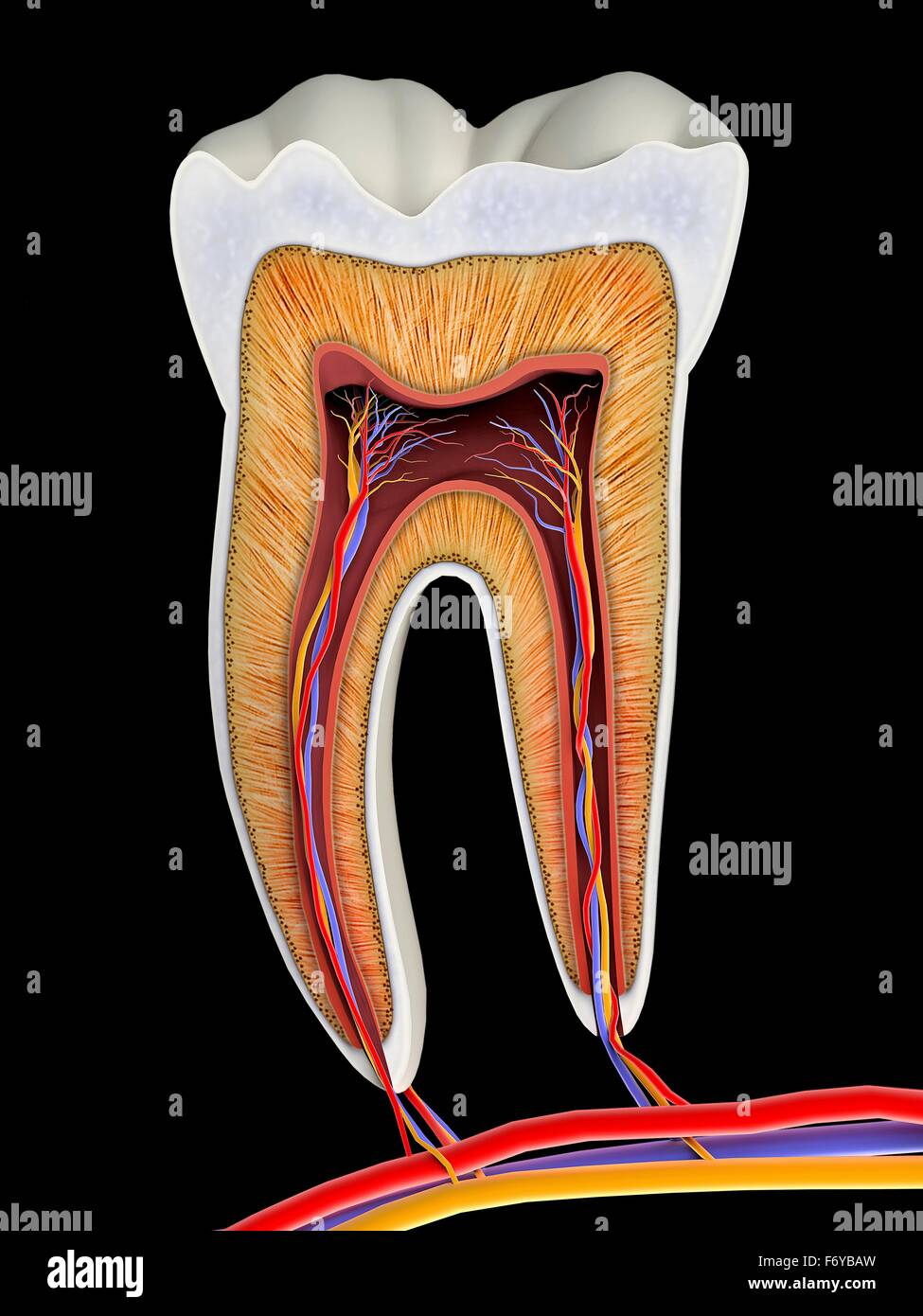
From wikimedia commons, the free media repository. Explaned distal and proximal epiphysis. Related posts of cross section of human bone diagram. Diagram of cross section of bone. Human tooth anatomy dentistry medical concept as a cross section of a molar with nerves and root canal symbol as a 3d related posts of cross section of human bone diagram bone anatomy of foot.
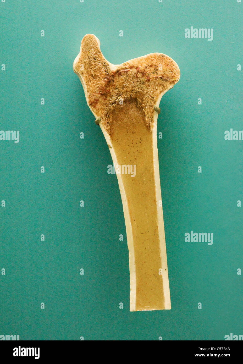
Cross Section Bone Human High Resolution Stock Photography And Images Alamy from c8.alamy.com Spongy bone diagram schematic diagram. Jump to navigation jump to search. They build the entire picture, improve your understanding, consolidate the information and facilitate recall. In a cross section of a bone we can see two types of bone tissue: Cross section through an model of an fracture in the neck of the femur (thigh bone). Diagram with articular cartilage, marrow, spongy bone, medullary cavity, endosteum, diaphysis, and periosteum. From wikimedia commons, the free media repository. This page discusses the calculation of cross section properties relevant to structural analysis, including centroid, moment of inertia, section modulus, and parallel axis theorem.
Metaphseal region on the left, diaphyseal region on the right.
Diagram with articular cartilage, marrow, spongy bone, medullary cavity, endosteum, diaphysis, and periosteum. Jump to navigation jump to search. How to draw the diagram of cross section of a leaf class x. Diagram of cross section of bone. Diagram with articular cartilage, marrow, spongy bone, medullary cavity, endosteum, diaphysis, and. Human tooth anatomy dentistry medical concept as a cross section of a molar with nerves and root canal symbol as a 3d related posts of cross section of human bone diagram bone anatomy of foot. □ on examining a cross section of any bone, it is composed of two kinds of bony tissue: For example, to read this diagram literally, since the cartilage can be seen inside the cutaway section of bone, it incorrectly indicates that the cartilage in fact goes through the bone structure, rather than just being found around the bone end. There are trabeculae in spongy bone which gives its sponge like appearance. □ the osteon consists of a system of bony lamellae arranged concentrically around a canal, which is called haversian canal. We don't draw the rest of the object, just the shape made when you cut through. Cross section through an model of an fracture in the neck of the femur (thigh bone). Metaphseal region on the left, diaphyseal region on the right.
How to draw the diagram of cross section of a leaf class x. The cross section of a rectangular pyramid is a rectangle. Spongy bone and compact bone. Spinal cord crosssection images stock photos vectors shutterstock. Select from premium cross section of bone images of the highest quality.
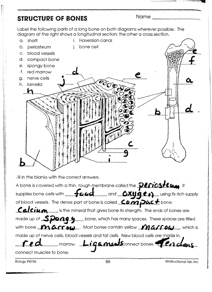
Diagram Frog Skeletal System Diagram Biology If8765 Answers Full Version Hd Quality If8765 Answers Diagramorama Casale Giancesare It from s3.studylib.net Explaned distal and proximal epiphysis. Diagram of a cross section of the coiled cochlea. Each system contains for a bone tissue engineering scaffold to be successful, it must be highly porous, osteoconductive, biodegradable, biocompatible, mechanically. Diagram with articular cartilage, marrow, spongy bone, medullary cavity, endosteum, diaphysis, and periosteum. These bone cells (described later) cause the bone to grow, repair, and remodel throughout life. Explaned distal and proximal epiphysis. In a cross section of a bone we can see two types of bone tissue: As shown in figure 2.
Metaphseal region on the left, diaphyseal region on the right. Bone tissue cross section diagram human oasissolutions co. The centroidal distance, c, is the distance from the centroid of a cross section to the extreme fiber. Under the moment m, its axis is bent into a circular curve, cross section mn and pq remain plane and normal to longitudinal lines (plane remains plane can be established by experimental result). A cross section of a human long bone. These bone cells (described later) cause the bone to grow, repair, and remodel throughout life. Cochlea diagram cross section as the travellers or messenger terminals are normally interconnected, the prevalent terminal is the only a single left. What are your bones made of? Diagram with articular cartilage, marrow, spongy bone, medullary cavity, endosteum, diaphysis, and periosteum. Bone marrow is the soft, highly vascular and flexible connective tissue within bone cavities which serve as the primary site of new blood cell production or bone marrow is the primary source of pluripotent stem cells that give rise to all hemopoietic cells (blood cells) including lymphocytes. We don't draw the rest of the object, just the shape made when you cut through. How to draw the diagram of cross section of a leaf class x. There are trabeculae in spongy bone which gives its sponge like appearance.
□ on examining a cross section of any bone, it is composed of two kinds of bony tissue: cross section of a bone. (micrograph provided by the regents of university of michigan.


0 Komentar